|
Vocapedia > Media > USA > NYT > Illustrations >
2008-2009

Loren Capelli
Letters
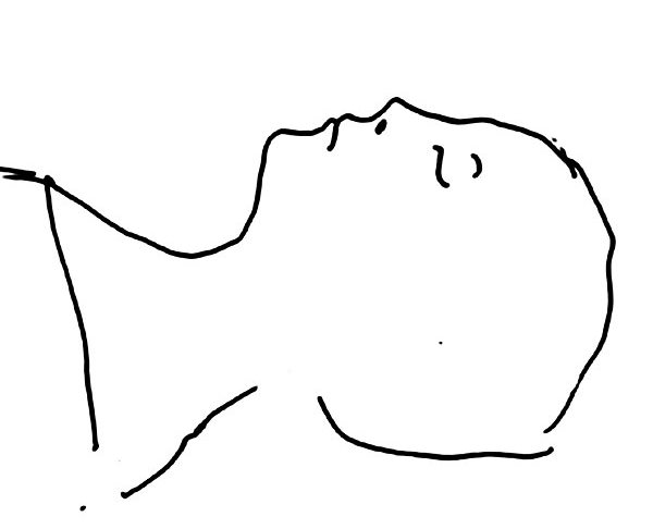
A Cadaver, for
the Sake of Science
April 2, 2009
The New York Times
To the Editor:
“Dead
Body of Knowledge,” by Christine Montross (Op-Ed, March 27), was a welcome
reminder of the value of human dissection.
An anatomical image is just an image. But the cadaver is the medical student’s
first patient and the first encounter with the emotional burden of becoming a
physician. The impact is nowhere more apparent than in the student’s initial
reaction to dissection.
The sight and smell of the body, the sounds of cutting and sawing, and the feel
of human flesh have effects both empathic and repulsive. These sensations
provide the earliest opportunity to examine how doctors manage (or mismanage)
the inevitable emotions associated with patient care.
Taught properly, dissection of the cadaver allows students to examine
themselves. Gary J. Kennedy
Bronx, March 30, 2009
The writer, a professor of psychiatry at the Albert Einstein College of
Medicine, is the co-author of “Cadaver Conference: A Psychiatrist in the Gross
Anatomy Course.”
•
To the Editor:
In my human anatomy class, I came to appreciate the uniqueness of the individual
human body. I found that structures like arteries, veins, ducts and even organs
vary greatly from what is shown in textbooks or on computer scans.
Dissecting the human cadaver proved to be a truly visceral experience. Seeing,
touching, smelling, moving muscles and bones helped me as an anatomy student
(and later as an anatomy instructor) to use a very important organ, my brain, to
decipher the complexity and beauty of the human body, both on an intellectual
and an emotional level.
I agree with Christine Montross. Cadaver dissection is indeed a vital part of
the anatomy curriculum.
Ginger Nathanson
Long Valley, N.J., March 28, 2009
•
To the Editor:
No research demonstrates that learning anatomy using medical imaging is inferior
to the information gained through the brief dissection of the one cadaver
allotted to each medical student.
The role of technology and medical imaging will inevitably increase in anatomy
courses. New digital tools like the ones we are developing at Stanford have been
shown to greatly enhance the learning experience. We now have the ability to
visualize and interact with anatomical information that was previously
inaccessible.
Teaching anatomy cannot be couched in an either-or framework; instead,
technology and cadavers should enhance each other. Only well-designed, validated
studies will provide answers to the issues introduced by Christine Montross.
W. Paul Brown
Stanford, Calif., March 30, 2009
The writer, a dentist, is a consulting associate professor in the Division of
Anatomy at Stanford University.
•
To the Editor:
I am disappointed to read in Christine Montross’s article that medical schools
are contemplating the transfer of anatomy from actual cadavers to virtual
reality because of cost.
As a first-year medical student, I find that most of our time is spent
relentlessly memorizing the exhaustive body of knowledge that has accumulated
through the years of research and science. It would be a great disservice to the
future of medicine to remove the single most important tool first- and
second-year medical students can physically access.
The cadavers present information that is simply inaccessible through the
computer. Could we simulate the systemic spread, consistency and color of the
many cancers discovered in the bodies? Would we empathize in the same way
looking through a monitor? Would a three-dimensional view be fully reproduced on
a two-dimensional screen?
It is my view that the answer to these, and countless more, is no.
Locke Uppendahl
Kansas City, Kan., March 27, 2009
To the Editor:
Most important, cadaver dissection enables students to appreciate the enormous
amount of anatomical variation among human beings. A computer program cannot
convey the fact that physical phenomena like aberrant arteries, accessory
glandular tissue or atrophied muscles belong to a particular formerly living
person.
Dissection allows students to recognize people as truly unique individuals in
body and in spirit, each with their own “irregularities.”
Without going into the lab, students may fail to consider a fundamental tenet of
medicine: no two patients are identical, and therefore all medical care must be
individualized. Geoff Rubin
New York, March 27, 2009
The writer is a first-year medical student at the Columbia College of Physicians
and Surgeons.
•
To the Editor:
Christine Montross has perfectly described the value and beauty of the human
body when compared with electronic imaging. In pathology we often say “a picture
is worth a thousand words, but a specimen is worth a thousand pictures.”
Dennis G. O’Neill
Manchester, Conn., March 27, 2009
The writer, a medical doctor, is director of the department of pathology and
laboratory services at Manchester Memorial Hospital.
A Cadaver, for the Sake
of Science, NYT, 2.4.2009,
http://www.nytimes.com/2009/04/02/opinion/l02cadaver.html
|